

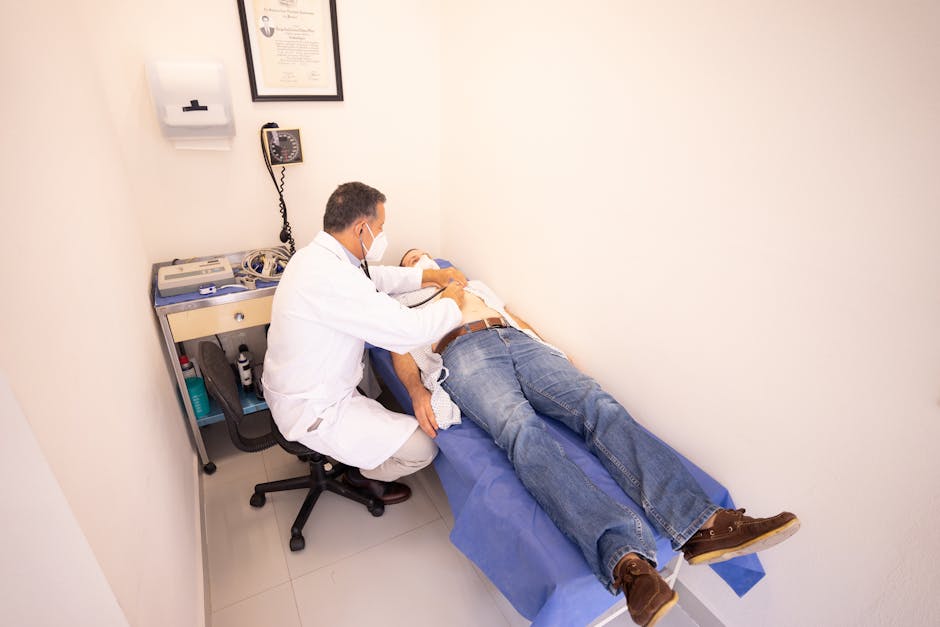
A normal X-ray after a car crash is reassuring and frequently misleading. It answers one question well, whether a bone is broken, and it is close to silent on the structures that actually generate months of pain afterward.
Understanding what each imaging modality can and cannot show is the difference between a diagnosis and a guess.
What Plain Radiography Actually Rules Out
Radiographs image bone density. They demonstrate fracture, dislocation, gross alignment and degenerative change. In the emergency setting that is exactly the right first question, because a missed cervical fracture is catastrophic and a missed ligament sprain is not.
What radiographs cannot show is the intervertebral disc, the spinal cord and nerve roots, ligaments, or muscle. A patient can have a completely normal cervical series and a substantial disc herniation on the same day.
Flexion and extension views add a functional dimension by imaging the spine in motion, which can suggest instability that a neutral film hides. This remains an indirect inference about ligament integrity rather than a direct look at it.
Where Magnetic Resonance Imaging Changes the Diagnosis
MRI images soft tissue directly. Disc herniation and protrusion, nerve root compression, ligamentous injury, spinal cord signal change and marrow edema all become visible.
Timing matters. Some findings are most apparent in the weeks following injury, while others evolve. A patient imaged the night of a collision and a patient imaged six weeks later can produce meaningfully different studies from the same underlying injury.
The interpretive challenge is that degenerative findings are extremely common in asymptomatic adults. Disc bulges and facet arthrosis appear in people with no pain at all, which means an MRI finding has to be correlated with the clinical examination rather than read in isolation.
Why Clinical Correlation Decides the Treatment Plan
An imaging report is a description of anatomy. It is not a diagnosis on its own, and it is not a treatment plan.
The finding that matters is the one that explains the patient's symptom pattern. A right-sided C6 radiculopathy on examination, matched to a right paracentral disc protrusion at C5-C6 on imaging, is a coherent picture. The same imaging finding in a patient with diffuse bilateral aching is a much weaker inference.
Practices built around injury care tend to keep imaging close to the clinical examination for this reason. Novaré Injury Care and Rehab, a medically integrated injury clinic in Fort Myers and Lehigh Acres, Florida, runs diagnostic imaging alongside its chiropractic and rehabilitation services so that findings inform the treatment plan directly rather than arriving through a separate referral weeks later.
How Imaging Supports the Documentation Record
Patients recovering from a collision are often managing an insurance claim at the same time as their symptoms. Objective imaging findings, correlated with a documented clinical examination, establish a record that a symptom diary cannot.
This is not a reason to image every patient. It is a reason to image deliberately, with a specific clinical question, and to make sure the report is read against the examination rather than filed on its own.
The useful sequence rarely changes. Examine first, form a hypothesis, then select the study that can confirm or refute it.
