

Your doctor just told you that you need an echocardiogram, and the first question that comes to mind is probably simple: what exactly happens next? Maybe you've had chest discomfort, an irregular heartbeat, or a routine visit that turned up something worth a closer look. Whatever brought you here, the uncertainty around a new test is often more stressful than the test itself.
An echocardiogram is one of the most common ways cardiologists evaluate how your heart is actually functioning, not just how it sounds through a stethoscope. It builds on the same warning signs we discuss in our guide to coronary artery disease risk factors and early warning signs, since chest pain, shortness of breath, and unexplained fatigue are frequently what prompt a doctor to order this exact test.
At BioSyntrx Medical Center, our cardiology team, part of a group of more than 100 physicians across eight specialties, orders echocardiograms every week for patients across the San Francisco Bay Area. We've found that most of the anxiety around the test comes from simply not knowing what to expect. This guide walks through the process step by step, what the images can and cannot show, and what typically happens after your results come back.
What Is an Echocardiogram?
An echocardiogram is an ultrasound test that uses high-frequency sound waves to create a moving picture of your heart's chambers, valves, and muscle. According to Mayo Clinic, the test lets a cardiologist watch the heart beating in real time and measure how much blood moves with each contraction, information a standard EKG or blood panel can't provide on its own. Unlike an X-ray, it uses no radiation, which makes it a test we can repeat as often as clinically necessary without added risk.

The word itself combines "echo," referring to sound waves bouncing back to form the image, with "cardiogram," a recording of the heart. Patients sometimes confuse an echocardiogram with an EKG, which measures electrical activity rather than physical structure. The two tests are complementary. An EKG tells us about rhythm. An echocardiogram tells us about structure and function, and together they give a far more accurate diagnosis than either one alone.
How an Echocardiogram Works
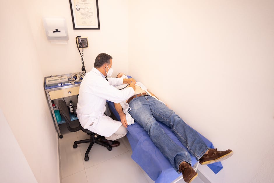
A standard transthoracic echocardiogram, the most common version, takes about 30 to 45 minutes. You'll lie on your left side on an exam table while a sonographer applies a small amount of gel to your chest and moves a handheld transducer across different points between your ribs. The transducer sends sound waves into your chest and captures the echoes as they bounce off your heart's structures, and a computer translates that data into a live image on the screen.
You may be asked to hold your breath briefly or shift position so the sonographer can get a clearer angle. It doesn't hurt. Most patients describe mild pressure from the probe and nothing more. In our clinic, a cardiologist typically reviews the images the same day, though a formal written report often takes 24 to 48 hours to reach your chart.
Will My Breasts Be Exposed During an Echocardiogram?
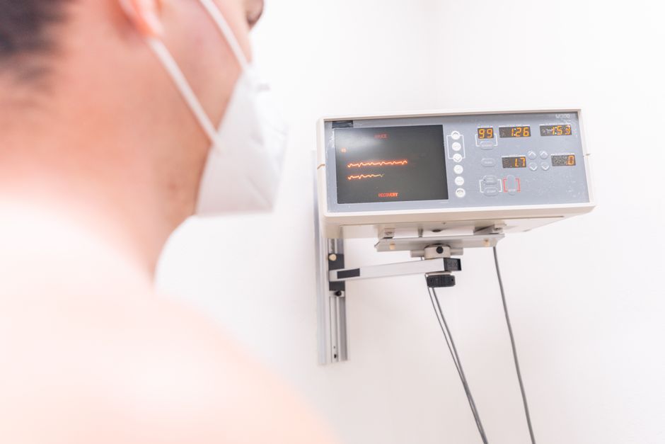
You'll change into a gown, and the technologist only uncovers the area of your chest actively being imaged, using a drape to keep the rest covered rather than leaving you exposed for the length of the test. This is standard practice for every patient, and you're welcome to request a chaperone if that helps you feel more comfortable.
How an echocardiogram is done on a woman is functionally identical to how it's done on a man. Breast tissue can occasionally make it a little harder to get a clean window of the heart, so the sonographer may press the probe more firmly or ask you to raise your arm above your head to shift tissue out of the way. It's a technical adjustment, not a different procedure. A female sonographer is generally available on request, and it's reasonable to ask for one when you schedule.
What Should You Not Do Before an Echocardiogram?
Avoid caffeine, nicotine, and heavy meals for a few hours before a standard echocardiogram, since both caffeine and nicotine can temporarily raise your heart rate and blood pressure, making the images harder to interpret accurately. For most transthoracic echocardiograms, you don't need to fast or stop your regular medications unless your doctor tells you otherwise.
A few simple habits make the appointment go more smoothly:
- Skip caffeine, including coffee, energy drinks, and chocolate, for at least 3 to 4 hours beforehand
- Avoid smoking or vaping the morning of your test
- Wear a two-piece outfit so you can change into a gown easily
- Leave lotion or oil off your chest, since it can interfere with the transducer's contact with your skin
- Bring a current medication list in case the sonographer or cardiologist has questions
- Arrive 10 to 15 minutes early to complete any updated intake paperwork
If your doctor has ordered a stress echocardiogram instead, the instructions change. You'll typically need to avoid eating for several hours beforehand and may need to hold certain heart medications, so always confirm the specific prep instructions for your test type when you schedule.
What an Echocardiogram Checks For
Cardiologists order echocardiograms to evaluate a wide range of structural and functional concerns, and the same test is useful whether you're being screened proactively or followed for a known condition. In our practice, we've caught problems in patients who had no symptoms at all, which is exactly why early evaluation matters so much in cardiology.
- Heart valve function, including narrowing (stenosis) or leaking (regurgitation)
- Pumping strength, measured as ejection fraction
- Chamber size and wall thickness, which can indicate long-term high blood pressure or prior strain
- Fluid buildup around the heart, known as pericardial effusion, which in severe cases can progress to cardiac tamponade
- Blood clots inside the heart's chambers
- Congenital heart defects present since birth
- Signs of a prior heart attack or damaged muscle tissue
- Irregular rhythms that may be linked to underlying structural changes
Because rhythm and structure are often connected, we frequently discuss both together with patients, which is why our guide to atrial fibrillation symptoms and irregular heartbeats pairs well with this one if you've noticed fluttering, skipping, or racing in your chest.
Does an Echocardiogram Show if Your Arteries Are Clogged?
An echocardiogram does not directly visualize the coronary arteries, so it cannot show blockages the way a coronary angiogram or CT angiogram can. What it can show is a downstream effect of blocked arteries, such as a section of heart muscle moving abnormally because it isn't getting enough blood flow, which often prompts a cardiologist to order more targeted testing.
"An echocardiogram uses sound waves to create moving images of your heart, showing its size, structure, and how well the chambers and valves are working, though it is not designed to directly image the coronary arteries themselves."
If your echocardiogram suggests reduced blood flow, the next step is often a stress test, a coronary calcium score, or a cardiac catheterization, depending on your symptoms and risk profile. We covered the risk side of this equation in our guide to cholesterol levels and heart disease risk, since elevated LDL cholesterol is one of the more common drivers of the plaque buildup an echocardiogram can only suggest, not confirm. It's also fair to be direct about the alternatives: for patients with a strong family history of coronary disease or ongoing chest pain despite a normal echocardiogram, a stress test or CT angiogram may be the more appropriate next step rather than simply repeating the echocardiogram.
If Echo Is Normal, Is My Heart Ok?
A normal echocardiogram is reassuring, but it only confirms that your heart's structure and pumping function looked normal at the moment of testing. It doesn't mean you're free of all cardiovascular risk going forward. Blood pressure, cholesterol, blood sugar, and coronary artery blockages can all exist independently of what an echocardiogram measures.

Most patients get a preliminary read the same day and a final written report within one to two business days. If everything looks normal and you were tested for a specific symptom like palpitations or shortness of breath, your cardiologist may still recommend monitoring, additional bloodwork, or a follow-up in six to twelve months depending on your risk factors. That's not a sign something was missed. It's simply how ongoing, evidence-based care works, and it lines up with the approach we describe in our guide to high blood pressure treatment and hypertension management for patients managing risk factors that don't always show up on a single test.
What Happens If They Find Something on an Echocardiogram?
If your echocardiogram shows an abnormality, your cardiologist explains what was found, how significant it is, and what comes next, ranging from watchful monitoring for a mild valve issue to urgent referral for something like a large pericardial effusion. The plan depends entirely on what's found and how it fits with your symptoms.
Some findings are minor and call for nothing more than periodic monitoring, such as mild valve regurgitation, which many adults have without ever noticing symptoms. Others, like significantly reduced ejection fraction or fluid building up around the heart, call for prompt follow-up and sometimes same-week referral to a specialist.
"Heart disease remains the leading cause of death for most groups in the United States, which is a major reason diagnostic tools like echocardiography play such a central role in early detection and prevention."
— CDC
Clear communication matters most at this stage. Board-certified providers recommend that patients ask direct questions about severity, next steps, and timeline rather than leaving an appointment with more anxiety than answers, and our team makes a point of walking through results in plain language instead of handing over a report full of unfamiliar terms.
Getting Ready for Your Echocardiogram: Practical Tips
Clinical educator Jean Terry, who works with our cardiology team on patient education, often reminds patients that a little preparation makes the whole experience faster and less stressful.
- Confirm which type of echocardiogram you're having, since stress and transesophageal versions carry different prep requirements
- Write down your symptoms and when they occur so you can describe them clearly to the sonographer
- Bring your insurance card and a photo ID to check-in
- Ask ahead of time whether your regular medications should be taken as scheduled
- Plan for a driver if you're having a stress or sedated version of the test
- Save detailed questions for your cardiologist, since the sonographer performing the test usually isn't the one who interprets the results
An echocardiogram is one of the more approachable tests in cardiology: no needles, no radiation, and typically no downtime, yet the information it provides shapes decisions about your heart health for years afterward. If your doctor has recommended one, or if you've been managing risk factors like high cholesterol or blood pressure without a recent look at how your heart is actually functioning, our cardiology team at BioSyntrx Medical Center is glad to walk you through scheduling and what to expect at every step of patient-focused, coordinated care.
